49 year old male with history of hypertension and hyerlipidemia that presents with hemoptysis for 1 week duration (about 1/4 a cup a day)
On admission vitals and labs were normal other than hypoxia on 87% that improved to 93% on 2 liters of oxygen
Chest X ray was normal on admission
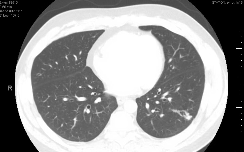
CT scan of the chest was peformed with the image below:
Flexable bronchoscopy was performed and revealed no source of bleeding or any endobronchial lesions
2d echo was ordered with bubble study that revealed a a right to left shunt with less than 10 bubbles seen crossing to the left atrium (after 4 cardiac cycles)
Shunt fraction was calculated to be 12%
Patient was diagnosed with Pulmonary AVM and was send to IR embolization
After the procedure patient had resolution of his symptoms with saturation improving to 97% on room air